Application of Bioluminescence Technology in Life Science
With the widespread use of luminescence technology in a variety of biological experiments, bioluminescence technology has increasingly become the preferred biodetection tool. In this article, we will discuss in detail the application of bioluminescence in biodetection and its advantages over other luminescence assays.
1 Characteristics of bioluminescence
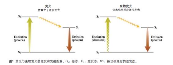
According to the different energy sources of photons, luminescence can be divided into the following four categories, one is fluorescence: relying on photon luminescence; the second is chemiluminescence: relying on chemical energy to illuminate; the third is radiance: relying on radioactive energy to emit light The fourth is electric energy illuminating: relying on electric energy to illuminate. Among them, bioluminescence is a luminescence that relies on the energy generated by natural enzymatic chemical reactions. It is a chemiluminescence, which naturally exists in many organisms, such as fireflies, jellyfish in the deep sea, and some bacteria. At present, the most widely used in life science research is fluorescence and chemiluminescence. As an important category of chemiluminescence, bioluminescence has also received increasing attention and application. The mechanism of matter luminescence is that the molecule is first excited, absorbing energy, transitioning from a stable ground state to an unstable excited state, and when it returns from the excited state to the ground state, energy is released in the form of photons (Fig. 1). The presence of luminescent molecules can be determined by detecting the emitted photons. The range of applications of luminescence technology in bioassays and their applications depends to a large extent on the source of energy for the excitation molecules. Because the source of the excitation energy is different, the relative signal strength (the relative value of the signal to the background) produced is different, and the sensitivity requirements for the detector are also different. Taking bioluminescence and fluorescence as an example: for fluorescence, on the one hand, due to the high efficiency of photon energy absorption of fluorescence, molecules can release a large amount of light energy, thereby generating a high-intensity optical signal for detection; on the other hand, High-flow excitation photons sometimes seriously interfere with the ability of the photodetector to emit photons, and may also excite the fluorophores contained in the biological samples themselves, resulting in other non-specific emission light, which leads to an excessively high experimental background. It is also because of this characteristic of fluorescence that the high-intensity signal in the reporter group coexists with the high-intensity experimental background, so that its ability to detect relative signal intensity is greatly reduced. For bioluminescence, it is in sharp contrast with fluorescence. Because bioluminescence relies on natural enzymatic chemical reactions to emit light, there is no need to introduce external light energy. Therefore, the experimental background of the reporter group using bioluminescence as a detection means is very bottom. Although the signal intensity of bioluminescence is not as high as that of fluorescence, bioluminescence can be one hundred or even more than one thousand times more sensitive than fluorescence due to its unique luminescence principle [1, 2]. This is one of the reasons why bioluminescence is becoming the preferred biological detection method in many biological research fields.
In practical applications, it is necessary to repeatedly weigh the requirements of signal strength and the influence of the experimental background to select the best detection technology. In the case of low photon detection capability, the experimental background is largely determined by the object of the instrument detection, such as microscopy and flow cytometry. Because of the need to detect changes in the optical signal of a single cell, the intensity of the emitted light signal is very high. High, therefore, fluorescence is often the first choice for this type of application. However, when a large number of biological samples need to be detected, the requirements for photon detection capability are correspondingly reduced, and the requirements for reducing the experimental background are correspondingly increased, such as in the detection of individual test tube samples, porous culture plate samples, or on organs ( Bioluminescence is popular for its low background and high sensitivity when performing image analysis. Moreover, since the bioluminescence detector does not require the use of an optical filter, it is possible to shorten the distance between the sample and the detector, thereby further improving the efficiency of detection. The sensitivity of bioluminescence can reach 10-20 mol, which is equivalent to more than 6,000 luciferase molecules. For a biological sample with hundreds of cells, a few molecules of luciferase can be detected in a single cell. Moreover, the high sensitivity of bioluminescence makes it possible to detect a wide range of concentrations, most of which can span 6-8 orders of magnitude, and the update of modern bioluminescence detector technology makes such a large span detection possible. In addition, since bioluminescence is derived from an enzymatic chemiluminescence reaction in a natural organism, when it is introduced into a biological sample, it does not add too much negative influence to normal biological activity. Moreover, since the substrate of the luciferase is encapsulated inside the luciferase molecule during the reaction, the normal enzymatic luminescence reaction can be protected from the outside interference to the utmost extent.

2 Bioluminescence as a reporter gene and its advantages
The most widely used bioluminescence in life science research is as a reporter gene for monitoring gene transcription activities. Among them, the naturally occurring luciferase in fireflies is usually the first choice of researchers. Firefly luciferase is a 61 kDa monomeric protein that catalyzes a luciferase substrate called luciferin, producing yellow-green light in the presence of ATP and oxygen ( The emitted light wavelength is 560 nm).
When using reporter genes to monitor gene transcriptional activity, the biggest concern of researchers is that the introduction of exogenous reporter genes may affect normal biological activity, especially when reporter genes often need to be linked to the molecular activities of their own cells in the organism. High concentrations of exogenous genes can over-emphasize the burden of biological cell activity, thereby affecting normal physiological activities and even causing abnormal physiological phenomena. Therefore, minimizing the concentration of reporter genes is the key to successful research. Taking green fluorescent protein as an example, it is a kind of naturally occurring luminescent protein. Because it can generate high-intensity emission fluorescence, it can obtain very clear and vivid microscopic images, which have been widely used in single-cell biological image analysis. . However, because the production of clear images usually requires high concentration of green fluorescent protein, which increases the burden of cellular activity inside the organism, making green fluorescent protein unsuitable as a reporter gene for monitoring gene transcription activities. So when choosing the right reporter gene, especially after comparison with green fluorescent protein, bioluminescence makes it stand out among many choices due to its low expression and high sensitivity.
Monitoring gene transcriptional activity requires that the reporter gene be able to rapidly monitor subtle dynamic changes in the target gene. The concentration of intracellular reporter genes depends on two major dynamic processes: the process of protein synthesis (regulated by the transcriptional activity of the gene) and the process of protein degradation. If the protein degradation process is slow, the protein will be highly stable, which will cause the background expression level of the protein to be too high, so the synthesis of new proteins produced by changes in gene transcriptional activity is not easily detected. As a result, the expression of the reporter gene does not accurately reflect changes in the transcriptional activity of the gene. Experiments have shown that protein degradation sequences such as mouse ornithine decarboxylase (ODC) are added to the luciferase sequence, which is rich in proline-glutamate-serine-threonine (proline- The sequence of glutamate-serinethreoninerich (PEST) can significantly improve the responsiveness of the detection technique and does not have much influence on the sensitivity of the detection means [3]. In contrast, due to the high expression and high stability of green fluorescent protein, the addition of protein degradation signals limits the sensitivity of detection to a large extent.
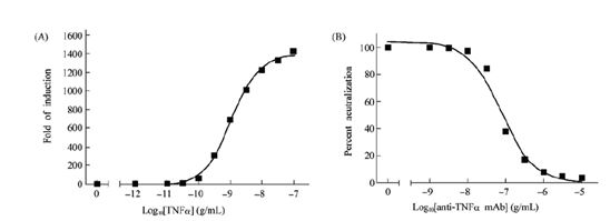
To further improve the efficiency of the detection of the gene, we optimized the codon of the luciferase gene sequence to increase its expression level in a variety of mammalian cells by 5 to 10 times. At the same time, in order to reduce the non-specificity of the gene. In regulation, we also optimized the luciferase vector to remove the conserved sequence of the mammalian transcription factor binding sequence on the vector, thereby greatly reducing the experimental background and significantly increasing the relative signal intensity of the experiment. The optimized luciferase reporter gene can be successfully applied in various biological research fields, such as the study of tumor necrosis factor signal transduction. Tumor necrosis factor (TNF) is an active factor capable of tumor cell necrosis produced by mononuclear-macrophages. It can enhance the phagocytic and digestive functions of neutrophils, promote adhesion to the vascular endothelium and migrate out of blood vessels, and Activation of the transcription factor NF-B regulates the expression of various genes including IL-1 and GM-CSF. In HEK293 cells exogenously expressing the NF-B luciferase reporter gene, tumor necrosis factor stimulation was effective to increase luciferase expression by more than 1000-fold (Fig. 2A). The same assay can also successfully detect the blocking efficiency of tumor necrosis factor blockers on tumor necrosis factor bioactivity (IC50 = 0.77 nmol, Figure 2B).

3 Other applications of bioluminescence
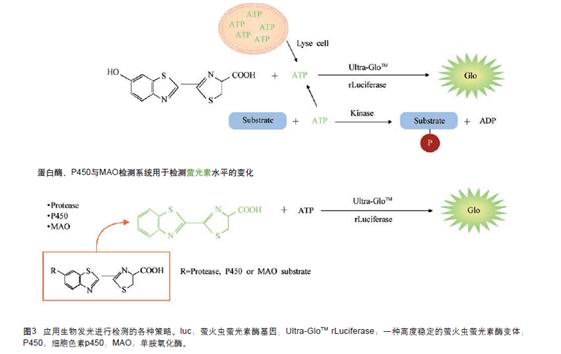
The intensity of bioluminescence depends on the concentration of each reactive component in the enzymatic chemical reaction, including luciferase concentration, ATP concentration, and concentration of luciferase substrate, luciferin. Generally, in a bioluminescence detection system, if the concentration of other components is kept excessive and constant, the concentration change of a specific component related to biological activity can be detected. The experiment we used to use luciferase as a reporter gene to monitor gene transcriptional activity is to directly correlate luciferase concentration with gene transcriptional activity. Luciferase concentration is our ultimate monitoring target. In addition, if we immobilize the luciferase concentration and make it excessive, bioluminescence can also be used to monitor biological activity associated with ATP or luciferin concentrations (Figure 3).
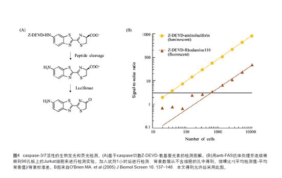
The principle of monitoring a biological activity by detecting the concentration of luciferin is to add a chemical group to the luciferin to modify a "modified" substrate that is not recognized by luciferase, but only through A specific biological reaction removes this chemical group, and luciferin can restore activity, and an enzymatic reaction can occur, so that we can link the intensity of bioluminescence to this specific biological response [1]. As shown in Figure 4, Asp-Glu-Val-Asp-6'-aminoluciferin is an inactive luciferin derivative, only when cysteine-aspartic protease (caspase-3) is present and excised The luciferase enzymatic reaction occurs when the sequence of the tetrapeptide releases free luciferin. Because cysteine-aspartic protease activity is an important indicator of apoptosis, this design makes bioluminescence an effective means of detecting apoptosis. Of course, this design can also be used to detect fluorescence apoptosis by adding the same tetrapeptide sequence to the fluorescent dye Rhodamine 110. However, due to the unique low expression and high sensitivity of bioluminescence, the use of bioluminescence to study apoptosis under the same experimental conditions is nearly one hundred times more sensitive than using fluorescence technology (Fig. 4). A similar design can also be used to detect the activity of other proteases using bioluminescence, such as caspase-8, caspase-9, dipeptidyl peptidase VI (DPPIV), caspase-3, calpain, proteosome Etc., and other enzymatic reactions such as CYP450 activity, monoamine oxidase, etc. can also be detected [2].
Bioluminescence was first used to detect the concentration of ATP and is still one of the most widely used methods for rapid detection of cell viability [4]. Compared with fluorescence-based cell viability assays (such as the use of tetrazolium), bioluminescence is used to detect the biological activity of mammalian cells with a sensitivity of more than one hundred times and only five minutes. Bioluminescence is used to detect the biological activity of bacteria, and the sensitivity is very high. When the number of bacteria is less than 10, it can be detected. The method of detecting ATP by bioluminescence can also be used to detect the concentration of a biological enzyme that consumes ATP. Taking kinases as an example, this method can be used as a universal means of detecting kinase activity because it can act on different phosphate receptors, including proteins, lipids and polysaccharide substrates. Recently, we used cAMP to regulate protein kinase A and protein kinase A kinase response to establish a bioluminescence assay for the detection of cAMP concentration changes in ATP consumption [5]. Specifically, the concentration of intracellular cAMP regulates the activity of protein kinase A, and activation of the protein kinase A catalyzes the corresponding phosphorylation reaction, which requires the consumption of ATP, so that the intracellular ATP concentration is reduced. Therefore, we detect The concentration of ATP reached is inversely proportional to the activity of protein kinase A and also inversely proportional to the concentration of cAMP. Since we are able to directly correlate bioluminescence with ATP concentrations, we have established a rapid and sensitive assay for the detection of G-protein-coupled receptor activity or phospholipidase activity in cells.

4 Conclusion
Bioluminescence has great potential and value in the study of complex biological organisms. Whether it emits a clear and quantitative signal at very low concentrations, or its low background in mammalian cells, bioluminescence technology has a unique position in revealing the mysteries of life. Therefore, bioluminescence detection technology will play an increasingly powerful role in many fields such as basic life science research and new drug development. (Bio Valley Bioon.com)

references:
1. O'Brien MA, Daily WJ, Hesselberth PE, Moravec RA, Scurria MA, Klaubert DH, Bulleit RF, Wood KV. Homogeneous, bioluminescent protease assays: caspase-3 as a model. J Biomol Screen , 2005,10(2 ): 137~148
2. Cali JJ, Ma D, Sobol M, Simpson DJ, Frackman S, Good TD, Daily WJ, Liu D. Luminogenic cytochrome P450 assays. Expert Opin Drug Metab Toxicol, 2006, 2(4): 629~645
3. Fan F, Wood KV. Bioluminescent assays for high-throughput screening. Assay Drug Dev Technol, 2007, 5(1): 127~136
4. Riss TL, Moravec RA. Use of multiple assay endpoints to investigate the effects of incubation time, dose of toxin, and plating density in cell based cytotoxicity assays. Assay Drug Dev Technol , 2004, 2(1): 51~62
5. Kumar M, Hsiao K, Vidugiriene J, Goueli SA. A bioluminescent-based, HTS-compatible assay to monitor G-protein-coupled receptor modulation of cellular cyclic AMP. Assay Drug Dev Technol, 2007, 5(2): 237 ~245
Clothing and accessories come in next after you choose the doll.
You can buy all kinds of clothes and wigs to dress up your doll and make her more sexy.
She can be a policeman, a nurse, a teacher, a call girl, a mysterious diviner, etc.
The most important thing is that she is the most Popular Sex Doll.
As there are multitudes of products you would seek from the seller, such as cleaner, heating rods ,wigs ,underwear ,lipstick ,
comb and so on as gifts random for dolls and others.


Sex Doll Full Body,Accessories Of Sex Dolls,Tpe Sex Dolls
Dongguan Chenkuang Biological Technology Co., Ltd , https://www.cksexdoll.com