Release date: 2017-03-15

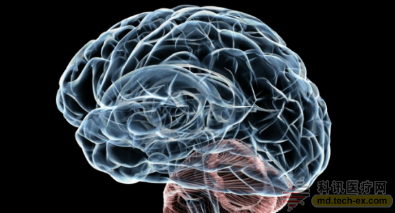
In-depth analysis of the human brain, we will find that every part of the brain has an amazing organizational structure. A large number of nerve bundles constitute a nerve conduction pathway, allowing nerve impulses to be accurately transmitted step by step. Neurons that are precisely distributed layer by layer within the cerebral cortex (grey) are tightly connected to each other to form a complex and precise neural network. Such an ordered structure indicates that the division and growth of each neuron are precisely regulated.
Once this regulatory mechanism is destroyed, the consequences will be terrible and will have a serious impact on the patient's cognitive and intellectual level.
In the rare genetic disease of tuberous sclerosis (TSC), there is a mutation in the TSC1 or TSC2 gene in patients with TSC. Proteins encoded by these two genes usually inhibit mTOR activity, and the mTOR signaling pathway is a key switch for many cell growth signals in neurons (and other cells).
If the TSC1 or TSC2 protein is not working properly, the continuously activated mTOR pathway will allow nerve cells to grow and divide. In this way, the brain will be occupied by these disorderly growth benign nodules, and the neural network will be destroyed.
Researchers at the Novartis Biomedical Research Center (NIBR) neuroscience research team have been trying to understand the intrinsic relationship between genetic mutations, abnormal cellular pathways, and disordered growth of nerve cells in the brain. To achieve this goal, they combined induced pluripotent stem cell technology with three-dimensional cell culture techniques to reprogram cells donated by TSC patients and healthy individuals into neurons and mimic the layered structure of cells in the cerebral cortex to form microclasses. The structure of the brain is like a "brain" in a petri dish.
This brain-like model was developed by NIBR neuroscientists Ajamete Kaykas and Max Salick using pluripotent stem cell induction techniques, originally from a healthy human somatic cell. In this model, a neuron-specific cytoskeletal protein β3 tubulin is stained red. In the figure, the more reddish the area, the more neurons are concentrated. At 14 days of growth, the neurons of this organ model have begun to stratify, as in the developing cerebral cortex.
Such an organ model allows Kaykas and Salick to study the development of nodular sclerosis in a natural three-dimensional structure similar to the cerebral cortex, and this model can also be used to test whether a potential treatment is effective, ie in a culture dish. Conduct an early efficacy trial. As a senior researcher in the NIBR Neuroscience Research Group, Kaykas said he was very excited to have the opportunity to study TSC and other diseases caused by abnormal activation of the mTOR pathway in a fully humanized system.
He said: "The technology of reducing human skin or blood cells to the state of embryonic stem cells and then inducing them into any other human cells, like "game rules changers", has greatly improved our construction of human disease models. Ability."
Author: Tom Ulrich Source: NERD Blog
Source: Novartis Group
Customized Drape Pack,Angiography Drape Pack,Disposable Surgical Drape Pack,Sterile Disposable Surgical Pack
ShaoXing SurgeCare Medical Products Co., Ltd. , https://www.sxsurgecaremedical.com